

Table of Contents
Toggle
Recent studies have uncovered intriguing connections between antiparasitic drugs and cancer treatment, with several medications showing promise in suppressing tumor growth through various mechanisms. As researchers continue to explore the potential of drug repurposing, antiparasitic agents like ivermectin, mebendazole, and others are emerging as candidates for novel cancer therapies.
Introduction to Antiparasitic Drugs in Cancer Treatment
Antiparasitic drugs, originally developed to treat parasitic infections, have recently emerged as potential anticancer agents. These drugs, including mebendazole, ivermectin, and fenbendazole, have shown promising antitumor properties in preclinical studies. The repurposing of these existing medications for cancer treatment has garnered significant attention due to their well-established safety profiles, low cost, and potential to target multiple cancer pathways.The anticancer mechanisms of antiparasitic drugs are diverse and not fully understood. However, studies suggest that they may inhibit key signaling pathways involved in cancer cell growth, survival, and metastasis. Additionally, some antiparasitic drugs have been shown to induce apoptosis, disrupt mitochondrial function, and enhance the efficacy of conventional chemotherapeutic agents.The repurposing of antiparasitic drugs for cancer treatment offers several advantages, including reduced development costs and faster clinical translation compared to novel drug discovery. As these drugs have already been approved for human use, their pharmacokinetic and toxicological profiles are well-characterized, potentially accelerating their application in oncology.
Mechanism of Action of Mebendazole on Cancer
Mebendazole (MBZ), a benzimidazole anthelmintic drug, has been repurposed for its anti-cancer properties. Originally approved by the FDA in 1971 for treating parasitic infections, MBZ has demonstrated efficacy in preclinical models of various cancers, including breast, ovarian, colorectal, and brain cancers.
The mechanisms by which MBZ exerts its anti-cancer effects are multifaceted and include tubulin disruption, induction of apoptosis, inhibition of angiogenesis, reduction of cancer stem cell properties, inhibition of hypoxia-inducible factors (HIFs), and cell cycle arrest.
The primary mechanism of MBZ’s anti-cancer action is the disruption of microtubule formation. MBZ binds to the colchicine-binding site of β-tubulin, preventing the polymerization of tubulin into microtubules. This disruption leads to mitotic arrest and apoptosis in cancer cells. Microtubules are essential for cell division, and their disruption halts the proliferation of cancer cells, leading to cell death (NCBI, 2023; DrugBank, 2023).
MBZ induces apoptosis through several pathways. It activates caspase-3 and caspase-9, which are crucial mediators of the apoptotic process. Additionally, MBZ inactivates BCL-2, an anti-apoptotic protein, thereby promoting cell death in cancer cells. This mechanism has been observed in various cancer cell lines, including melanoma and adrenocortical carcinoma (NCBI, 2023; ecancer, 2023).
Angiogenesis, the formation of new blood vessels, is critical for tumor growth and metastasis. MBZ inhibits angiogenesis by downregulating vascular endothelial growth factor (VEGF) and other angiogenic factors. This inhibition starves the tumor of necessary nutrients and oxygen, thereby inhibiting its growth and spread (NCBI, 2023; ecancer, 2023).
MBZ has been shown to reduce the stem-like properties of cancer cells, particularly in triple-negative breast cancer (TNBC). It decreases the expression of integrin β4 (ITGβ4), a protein associated with cancer stemness and metastasis. By targeting cancer stem cells, MBZ helps in reducing tumor recurrence and metastasis (Breast Cancer Research, 2022).
Hypoxia-inducible factors (HIFs) play a significant role in the adaptation of cancer cells to low oxygen conditions, promoting survival and chemoresistance. MBZ inhibits the transcriptional activity of HIF-1α, HIF-2α, and HIF-1β, thereby reducing the hypoxia-induced phenotype in cancer cells. This inhibition helps in sensitizing cancer cells to chemotherapy and reducing their metastatic potential (NCBI, 2023).
MBZ induces G2/M cell cycle arrest in cancer cells. This arrest prevents the cells from undergoing mitosis, leading to cell death. The G2/M checkpoint is crucial for ensuring that cells do not enter mitosis with damaged DNA, and its disruption by MBZ contributes to its cytotoxic effects on cancer cells (Breast Cancer Research, 2022).
MBZ has been shown to synergize with other chemotherapeutic agents and radiation therapy. It enhances the efficacy of these treatments by sensitizing cancer cells to their effects. For instance, MBZ has been observed to decrease the expression of multi-drug resistance proteins, thereby overcoming chemoresistance in cancer cells (ecancer, 2023).

Conclusion
Mebendazole’s multifaceted mechanisms of action make it a promising candidate for repurposing as an anti-cancer agent. Its ability to disrupt microtubules, induce apoptosis, inhibit angiogenesis, reduce cancer stem cell properties, inhibit HIFs, and cause cell cycle arrest, along with its synergistic effects with other therapies, highlight its potential in cancer treatment. Further clinical trials and studies are warranted to fully explore and validate its efficacy and safety in oncology.
Sources
- NCBI. (2023). Mebendazole Treatment Disrupts the Transcriptional Activity. Retrieved from https://www.ncbi.nlm.nih.gov/pmc/articles/PMC9954103/
- ecancer. (2023). Repurposing Drugs in Oncology (ReDO)—mebendazole as an anti-cancer agent. Retrieved from https://ecancer.org/en/journal/article/443-repurposing-drugs-in-oncology-redo-mebendazole-as-an-anti-cancer-agent
- Breast Cancer Research. (2022). Mebendazole prevents distant organ metastases in part by decreasing ITGβ4 expression and cancer stemness. Retrieved from https://breast-cancer-research.biomedcentral.com/articles/10.1186/s13058-022-01591-3
- ScienceDirect. (2020). Potential and mechanism of mebendazole for treatment and maintenance of ovarian cancer. Retrieved from https://www.sciencedirect.com/science/article/abs/pii/S009082582034018X
- DrugBank. (2023). Mebendazole: Uses, Interactions, Mechanism of Action. Retrieved from https://go.drugbank.com/drugs/DB00643
Mechanism of Action of Fenbendazole on Cancer
Fenbendazole (FZ), a benzimidazole anthelmintic drug primarily used in veterinary medicine, has shown potential as an anti-cancer agent through various mechanisms. These mechanisms include microtubule disruption, induction of apoptosis, cell cycle arrest, inhibition of glucose uptake, and modulation of multiple cellular pathways.
Fenbendazole acts as a moderate microtubule destabilizing agent. It binds to β-tubulin, preventing the polymerization of microtubules, which are essential for cell division. This disruption leads to mitotic arrest and apoptosis in cancer cells. The destabilization of microtubules interferes with the structural integrity and function of the mitotic spindle, crucial for chromosome segregation during cell division (Dogra, Kumar, & Mukhopadhyay, 2018).
Fenbendazole induces apoptosis through both p53-dependent and p53-independent pathways. In colorectal cancer (CRC) cells, fenbendazole increases p53 expression, leading to p53-mediated apoptosis. In 5-fluorouracil-resistant CRC cells, fenbendazole triggers apoptosis without affecting p53 expression and enhances ferroptosis by inhibiting the expression of GPX4 and SLC7A11 (Park et al., 2022).
Fenbendazole induces G2/M phase cell cycle arrest. This arrest prevents cells from undergoing mitosis, leading to cell death. The G2/M checkpoint ensures that cells do not enter mitosis with damaged DNA, and its disruption by fenbendazole contributes to its cytotoxic effects on cancer cells (Park et al., 2022).
Fenbendazole reduces glucose uptake in cancer cells by downregulating glucose transporters (GLUT) and key glycolytic enzymes. This reduction in glucose uptake starves cancer cells of the energy required for their rapid growth and proliferation, thereby inhibiting tumor growth (Dogra, Kumar, & Mukhopadhyay, 2018).
Fenbendazole exerts its antitumor effects by modulating various cellular pathways. It activates p53, a tumor suppressor protein, and modulates genes involved in multiple cellular pathways, leading to cell death. This pleiotropic effect makes fenbendazole a promising candidate for cancer therapy, as it targets multiple pathways involved in tumorigenesis (Dogra, Kumar, & Mukhopadhyay, 2018).
Fenbendazole has shown potential in combination with other chemotherapeutic agents. Although some studies have reported additive cytotoxicities when combined with drugs like docetaxel, the exact nature of these interactions requires further investigation (PMC, 2013).

Conclusion
Fenbendazole’s multifaceted mechanisms of action make it a promising candidate for repurposing as an anti-cancer agent. Its ability to disrupt microtubules, induce apoptosis, cause cell cycle arrest, inhibit glucose uptake, and modulate multiple cellular pathways highlights its potential in cancer treatment. Further clinical trials and studies are warranted to fully explore and validate its efficacy and safety in oncology.
Sources
- Dogra, N., Kumar, A., & Mukhopadhyay, T. (2018). Fenbendazole acts as a moderate microtubule destabilizing agent and causes cancer cell death by modulating multiple cellular pathways. Scientific Reports, 8, 11926. https://doi.org/10.1038/s41598-018-30158-6
- Park, D., Lee, J.-H., & Yoon, S.-P. (2022). Anti-cancer effects of fenbendazole on 5-fluorouracil-resistant colorectal cancer cells. PMC. https://www.ncbi.nlm.nih.gov/pmc/articles/PMC9437363/
- PMC. (2013). Fenbendazole as a Potential Anticancer Drug. NCBI. https://www.ncbi.nlm.nih.gov/pmc/articles/PMC3580766/
Mechanism of Action of Niclosamide on Cancer
Niclosamide, an FDA-approved anthelmintic drug, has shown significant potential as an anti-cancer agent. Its mechanisms of action in cancer treatment are diverse and involve the inhibition of multiple oncogenic pathways, induction of apoptosis, and disruption of cellular metabolism.
Niclosamide has been identified as a direct inhibitor of the signal transducer and activator of transcription 3 (STAT3) pathway. STAT3 is a critical transcription factor involved in cell growth and survival. By inhibiting STAT3, niclosamide suppresses the proliferation of cancer cells and induces apoptosis. This mechanism has been observed in various cancers, including esophageal, lung, and head and neck cancers (Spandidos Publications, 2019; NCBI, 2017).
Niclosamide acts as an uncoupling agent for oxidative phosphorylation in mitochondria. This action disrupts the energy production in cancer cells, leading to reduced ATP levels and increased reactive oxygen species (ROS) production. The mitochondrial dysfunction induced by niclosamide contributes to its cytotoxic effects on cancer cells (NCBI, 2017; DrugBank, 2023).
The Wnt/β-catenin signaling pathway is crucial for cell proliferation and differentiation. Niclosamide inhibits this pathway by downregulating β-catenin and other associated proteins. This inhibition leads to reduced cancer cell growth and metastasis. This mechanism has been particularly noted in colorectal, breast, and ovarian cancers (ScienceDirect, 2022; NCBI, 2017).
Niclosamide induces apoptosis through multiple pathways. It activates caspases, which are essential for the execution of apoptosis. Additionally, niclosamide modulates the expression of pro-apoptotic and anti-apoptotic proteins, tipping the balance towards cell death. This apoptotic effect has been observed in various cancer cell lines, including acute myeloid leukemia and hepatocellular carcinoma (Spandidos Publications, 2019; ScienceDirect, 2022).
The nuclear factor kappa-light-chain-enhancer of activated B cells (NF-κB) pathway is involved in inflammation and cell survival. Niclosamide inhibits the NF-κB pathway, leading to reduced expression of survival genes and increased sensitivity of cancer cells to apoptosis. This mechanism has been demonstrated in leukemia and glioma cells (NCBI, 2017; ScienceDirect, 2022).
The mammalian target of rapamycin (mTOR) pathway is a key regulator of cell growth and metabolism. Niclosamide inhibits mTOR signaling, leading to reduced protein synthesis and cell proliferation. This inhibition has been observed in breast and ovarian cancers (Spandidos Publications, 2019; NCBI, 2017).
Niclosamide has shown synergistic effects when combined with other chemotherapeutic agents. For example, it enhances the efficacy of paclitaxel in esophageal cancer and cisplatin in renal cell carcinoma. These combinations lead to improved therapeutic outcomes and reduced drug resistance (ScienceDirect, 2022; NCBI, 2017).

Conclusion
Niclosamide’s multifaceted mechanisms of action make it a promising candidate for repurposing as an anti-cancer agent. Its ability to inhibit multiple oncogenic pathways, induce apoptosis, disrupt cellular metabolism, and synergize with other therapies highlights its potential in cancer treatment. Further clinical trials and studies are warranted to fully explore and validate its efficacy and safety in oncology.
Sources
- Spandidos Publications. (2019). Niclosamide inhibits the cell proliferation and enhances the … Retrieved from https://www.spandidos-publications.com/10.3892/or.2019.7449
- NCBI. (2017). Niclosamide: Beyond an antihelminthic drug – PMC. Retrieved from https://www.ncbi.nlm.nih.gov/pmc/articles/PMC5628105/
- ScienceDirect. (2022). Combination of niclosamide and current therapies to overcome … Retrieved from https://www.sciencedirect.com/science/article/pii/S0753332222011787
- DrugBank. (2023). Niclosamide: Uses, Interactions, Mechanism of Action. Retrieved from https://go.drugbank.com/drugs/DB06803
- ScienceDirect. (2022). Niclosamide – an overview. Retrieved from https://www.sciencedirect.com/topics/pharmacology-toxicology-and-pharmaceutical-science/niclosamide
Mechanism of Action of Ivermectin on Cancer
Ivermectin, a well-known antiparasitic drug, has shown promising potential as an anti-cancer agent. Its mechanisms of action in cancer treatment are diverse and involve the inhibition of multiple oncogenic pathways, induction of apoptosis, and disruption of cellular metabolism.
Ivermectin effectively suppresses the proliferation and metastasis of cancer cells. It has been shown to inhibit the growth of colorectal cancer cell lines SW480 and SW1116 dose-dependently, promoting cell apoptosis and increasing Caspase-3/7 activity (NCBI, 2023). Additionally, ivermectin inhibits the proliferation of several tumor cells by regulating multiple signaling pathways, suggesting its potential as an anticancer drug (ScienceDirect, 2020).
Ivermectin induces apoptosis through various mechanisms. It promotes programmed cancer cell death, including apoptosis, autophagy, and pyroptosis. The drug induces apoptosis and autophagy in a mutually regulated manner, enhancing the sensitivity of cancer cells to chemotherapeutic drugs and reducing resistance (NCBI, 2020). In chronic myeloid leukemia, ivermectin selectively induces apoptosis by causing mitochondrial dysfunction (ScienceDirect, 2020).
Ivermectin induces mitochondrial dysfunction, leading to increased reactive oxygen species (ROS) production and cell death. This mechanism has been observed in glioblastoma, where ivermectin inhibits angiogenesis, growth, and survival of cancer cells by inducing mitochondrial dysfunction and oxidative stress (ScienceDirect, 2020).
The nuclear factor kappa-light-chain-enhancer of activated B cells (NF-κB) pathway is involved in inflammation and cell survival. Ivermectin inhibits the NF-κB pathway, leading to reduced expression of survival genes and increased sensitivity of cancer cells to apoptosis. This mechanism has been demonstrated in various cancer types, including glioma and leukemia (Nature, 2021).
The Wnt/β-catenin signaling pathway is crucial for cell proliferation and differentiation. Ivermectin inhibits this pathway by downregulating β-catenin and other associated proteins, leading to reduced cancer cell growth and metastasis. This inhibition has been particularly noted in colorectal and breast cancers (NCBI, 2020).
Ivermectin has been identified as an inhibitor of the signal transducer and activator of transcription 3 (STAT3) pathway. STAT3 is a critical transcription factor involved in cell growth and survival. By inhibiting STAT3, ivermectin suppresses the proliferation of cancer cells and induces apoptosis (NCBI, 2020).
Ivermectin has shown synergistic effects when combined with other chemotherapeutic agents. For example, it enhances the efficacy of cisplatin in epithelial ovarian cancer by suppressing the Akt/mTOR signaling pathway. These combinations lead to improved therapeutic outcomes and reduced drug resistance (ScienceDirect, 2020).

Conclusion
Ivermectin’s multifaceted mechanisms of action make it a promising candidate for repurposing as an anti-cancer agent. Its ability to inhibit multiple oncogenic pathways, induce apoptosis, disrupt mitochondrial function, and synergize with other therapies highlights its potential in cancer treatment. Further clinical trials and studies are warranted to fully explore and validate its efficacy and safety in oncology.
Sources
- NCBI. (2023). Outcome of Ivermectin in Cancer Treatment: An Experience in Loja. Retrieved from https://www.ncbi.nlm.nih.gov/pmc/articles/PMC10054244/
- ScienceDirect. (2020). Ivermectin, a potential anticancer drug derived from an antiparasitic. Retrieved from https://www.sciencedirect.com/science/article/abs/pii/S1043661820315152
- NCBI. (2020). Ivermectin, a potential anticancer drug derived from an antiparasitic. Retrieved from https://www.ncbi.nlm.nih.gov/pmc/articles/PMC7505114/
- Nature. (2021). The mechanisms of action of ivermectin against SARS-CoV-2. Retrieved from https://www.nature.com/articles/s41429-021-00491-6
- DrugBank. (2023). Ivermectin: Uses, Interactions, Mechanism of Action. Retrieved from https://go.drugbank.com/drugs/DB00602
Mechanism of Action of Albendazole on Cancer
Albendazole, a benzimidazole anthelmintic drug, has shown significant potential as an anti-cancer agent. Its mechanisms of action in cancer treatment are diverse and involve the inhibition of multiple oncogenic pathways, induction of apoptosis, and disruption of cellular metabolism.
Albendazole disrupts microtubule formation by binding to β-tubulin, preventing its polymerization into microtubules. This disruption leads to mitotic arrest and apoptosis in cancer cells. Microtubules are essential for cell division, and their disruption halts the proliferation of cancer cells, leading to cell death (NCBI, 2023; DrugBank, 2023).
Albendazole induces apoptosis through several pathways. It activates caspase-3 and caspase-9, which are crucial mediators of the apoptotic process. Additionally, albendazole inactivates BCL-2, an anti-apoptotic protein, thereby promoting cell death in cancer cells. This mechanism has been observed in various cancer cell lines, including gastric cancer (MDPI, 2021).
Albendazole has been identified as an inhibitor of the signal transducer and activator of transcription 3 (STAT3) and STAT5 pathways. These pathways are critical for cell growth and survival. By inhibiting STAT3 and STAT5, albendazole suppresses the proliferation of cancer cells and induces apoptosis. This mechanism has been particularly noted in gastric cancer cells (MDPI, 2021).
Albendazole disrupts cellular metabolism by inhibiting key metabolic pathways. It affects glucose uptake and utilization, leading to reduced energy production in cancer cells. This metabolic disruption contributes to the cytotoxic effects of albendazole on cancer cells (ScienceDirect, 2016).
Angiogenesis, the formation of new blood vessels, is critical for tumor growth and metastasis. Albendazole inhibits angiogenesis by downregulating vascular endothelial growth factor (VEGF) and other angiogenic factors. This inhibition starves the tumor of necessary nutrients and oxygen, thereby inhibiting its growth and spread (ScienceDirect, 2016).
Albendazole induces oxidative stress in cancer cells by increasing the production of reactive oxygen species (ROS). This oxidative stress leads to cellular damage and apoptosis. The increased ROS levels disrupt the redox balance in cancer cells, contributing to their death (NCBI, 2023).
Albendazole has shown synergistic effects when combined with other chemotherapeutic agents. For example, it enhances the efficacy of paclitaxel and cisplatin in various cancer models. These combinations lead to improved therapeutic outcomes and reduced drug resistance (ScienceDirect, 2016).

Conclusion
Albendazole’s multifaceted mechanisms of action make it a promising candidate for repurposing as an anti-cancer agent. Its ability to disrupt microtubules, induce apoptosis, inhibit STAT3 and STAT5 signaling, disrupt cellular metabolism, inhibit angiogenesis, induce oxidative stress, and synergize with other therapies highlights its potential in cancer treatment. Further clinical trials and studies are warranted to fully explore and validate its efficacy and safety in oncology.
Sources
- NCBI. (2023). Albendazole. Retrieved from https://www.ncbi.nlm.nih.gov/books/NBK553082/
- MDPI. (2021). Albendazole Exhibits Anti-Neoplastic Actions against Gastric Cancer Cells by Affecting STAT3 and STAT5 Activation by Pleiotropic Mechanism(s). Retrieved from https://www.mdpi.com/2227-9059/9/4/362
- DrugBank. (2023). Albendazole: Uses, Interactions, Mechanism of Action. Retrieved from https://go.drugbank.com/drugs/DB00518
- ScienceDirect. (2016). Albendazole as a promising molecule for tumor control. Retrieved from https://www.sciencedirect.com/science/article/pii/S2213231716301926
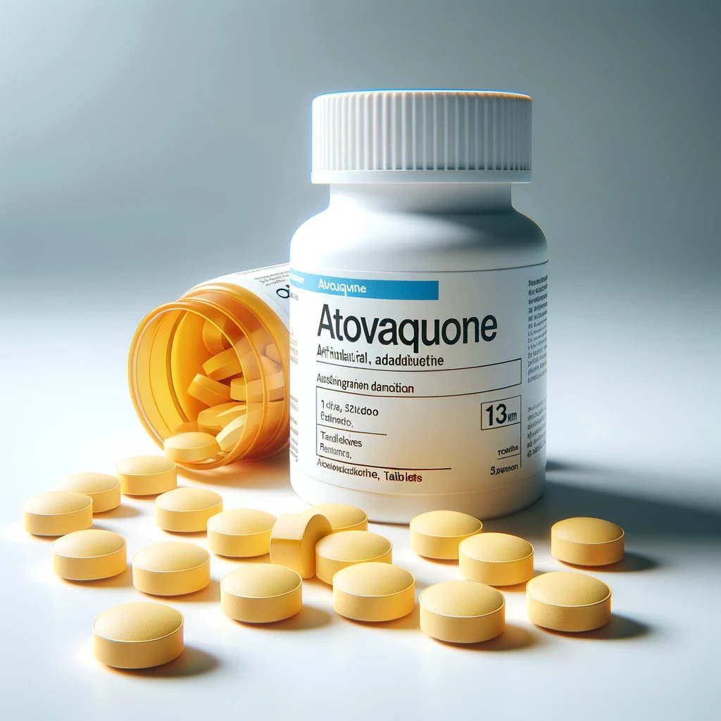
Mechanism of Action of Atovaquone on Cancer
Atovaquone, an FDA-approved anti-malarial drug, has shown significant potential as an anti-cancer agent. Its mechanisms of action in cancer treatment are diverse and involve the inhibition of multiple oncogenic pathways, induction of apoptosis, disruption of cellular metabolism, and modulation of the tumor microenvironment.
Atovaquone primarily exerts its anti-cancer effects by inhibiting oxidative phosphorylation in the mitochondria. It inhibits the electron transport chain (ETC) complex III, leading to decreased oxygen consumption and ATP production in cancer cells. This disruption of energy metabolism induces oxidative stress and apoptosis in cancer cells (PubMed, 2023; NCBI, 2023).
By inhibiting complex III of the ETC, atovaquone increases the production of mitochondrial reactive oxygen species (mROS) in cancer cells. The resulting oxidative stress depletes intracellular glutathione (GSH) levels, leading to cellular damage and apoptosis. This mechanism has been observed in various cancer cell lines, including ovarian and breast cancers (NCBI, 2023; Nature, 2020).
Atovaquone has been shown to inhibit the transcriptional activity of hypoxia-inducible factors (HIFs), such as HIF-1α, HIF-2α, and HIF-1β. HIFs play a crucial role in the adaptation of cancer cells to low oxygen conditions, promoting survival and chemoresistance. By inhibiting HIFs, atovaquone sensitizes cancer cells to chemotherapy and reduces their metastatic potential (Nature, 2023; Frontiers in Pharmacology, 2022).
Atovaquone inhibits Na+/K+-ATPase activity, which is essential for maintaining cellular ion balance and membrane potential. This inhibition leads to increased intracellular sodium levels, disrupting cellular homeostasis and inducing apoptosis. The oxidative stress caused by atovaquone further degrades Na+/K+-ATPase, contributing to its cytotoxic effects on cancer cells (Veterinary World, 2023; NCBI, 2023).
Atovaquone modulates the tumor microenvironment by reducing hypoxia and enhancing the immune response. It alleviates tumor hypoxia by inhibiting oxygen consumption, which can improve the efficacy of immunotherapies. For example, atovaquone has been shown to enhance the anti-tumor effects of PD-L1 inhibitors by promoting the activation of CD8+ T cells, leading to a stronger immune-mediated response against tumors (Nature, 2020; Nature, 2023).
Atovaquone has demonstrated synergistic effects when combined with other chemotherapeutic agents and immunotherapies. For instance, it enhances the efficacy of platinum-based chemotherapies (cisplatin, carboplatin) by increasing oxidative stress in cancer cells. Additionally, atovaquone has been shown to potentiate the effects of PD-L1 inhibitors, leading to improved therapeutic outcomes and reduced drug resistance (Nature, 2020; NCBI, 2023).
Conclusion
Atovaquone’s multifaceted mechanisms of action make it a promising candidate for repurposing as an anti-cancer agent. Its ability to inhibit oxidative phosphorylation, induce oxidative stress, inhibit HIFs, modulate the tumor microenvironment, and synergize with other therapies highlights its potential in cancer treatment. Further clinical studies are warranted to fully explore and validate its efficacy and safety in oncology.
Sources
- PubMed. (2023). Atovaquone: An Inhibitor of Oxidative Phosphorylation as Studied in Gynecologic Cancers. Retrieved from https://pubmed.ncbi.nlm.nih.gov/35565426/
- NCBI. (2023). Atovaquone: An antiprotozoal drug, suppresses primary and resistant breast tumor growth by inhibiting HER2/β-catenin signaling. Retrieved from https://www.ncbi.nlm.nih.gov/pmc/articles/PMC6905100/
- Veterinary World. (2023). Atovaquone exerts its anticancer effect by inhibiting Na+/K+-ATPase. Retrieved from https://www.veterinaryworld.org/Vol.16/June-2023/1.pdf
- Nature. (2020). The anti-malarial drug atovaquone potentiates platinum-mediated cancer cell death by increasing oxidative stress. Retrieved from https://www.nature.com/articles/s41420-020-00343-6
- Nature. (2023). Antitumour effect of the mitochondrial complex III inhibitor atovaquone. Retrieved from https://www.nature.com/articles/s41419-023-06405-8
Frontiers in Pharmacology. (2022). Targeting hypoxia-inducible factors for breast cancer therapy. Retrieved from https://www.frontiersin.org/journals/pharmacology/articles/10.3389/fphar.2022.1064661/full
Mechanism of Action of Pyrantel Pamoate on Cancer
Pyrantel pamoate, an FDA-approved anthelmintic drug, has shown potential as an anti-cancer agent. Its mechanisms of action in cancer treatment are diverse and involve the inhibition of multiple oncogenic pathways, induction of apoptosis, and disruption of cellular metabolism.
Pyrantel pamoate (PP) has been shown to inhibit the WNT signaling pathway, which plays a crucial role in the self-renewal and proliferation of cancer stem cells (CSCs). By inhibiting WNT signaling, PP reduces the expression of β-catenin, a key protein in this pathway, leading to decreased tumor growth and metastasis. This mechanism has been observed in various cancer types, including breast, lung, and prostate cancers (NCBI, 2023; PLOS ONE, 2013).
PP disrupts mitochondrial function by inhibiting oxidative phosphorylation. This inhibition leads to decreased ATP production and increased production of reactive oxygen species (ROS), resulting in oxidative stress and apoptosis in cancer cells. The disruption of mitochondrial function is a critical mechanism by which PP exerts its anti-cancer effects (NCBI, 2023; ScienceDirect, 2021).
PP has been shown to target cancer stem cells (CSCs), which are responsible for tumor initiation, progression, and resistance to conventional therapies. By inhibiting the WNT pathway and mitochondrial function, PP impairs the self-renewal and survival of CSCs, leading to reduced tumor growth and metastasis. This effect has been demonstrated in multiple cancer types, including melanoma, leukemia, glioblastoma, and cancers of the prostate, pancreas, lung, ovary, and breast (NCBI, 2023; PLOS ONE, 2013).
PP induces apoptosis in cancer cells through various mechanisms. It activates caspases, which are essential for the execution of apoptosis. Additionally, PP modulates the expression of pro-apoptotic and anti-apoptotic proteins, tipping the balance towards cell death. This apoptotic effect has been observed in various cancer cell lines, including breast and lung cancers (NCBI, 2023; PLOS ONE, 2013).
PP has demonstrated significant anti-tumor activity in vivo. It inhibits primary and secondary tumor growth, reduces lung metastasis, and synergizes with radiotherapy. These effects are attributed to its ability to inhibit the WNT pathway, disrupt mitochondrial function, and target CSCs. The inhibition of tumor growth and metastasis by PP has been observed in various preclinical models (NCBI, 2023; PLOS ONE, 2013).

Conclusion
Pyrantel pamoate’s multifaceted mechanisms of action make it a promising candidate for repurposing as an anti-cancer agent. Its ability to inhibit the WNT signaling pathway, disrupt mitochondrial function, target cancer stem cells, induce apoptosis, and inhibit tumor growth and metastasis highlights its potential in cancer treatment. Further clinical studies are warranted to fully explore and validate its efficacy and safety in oncology.
Sources
- NCBI. (2023). Pyrvinium Pamoate: Past, Present, and Future as an Anti-Cancer Drug. Retrieved from https://www.ncbi.nlm.nih.gov/pmc/articles/PMC9775650/
- ScienceDirect. (2021). Anthelmintics for drug repurposing: Opportunities and challenges. Retrieved from https://www.sciencedirect.com/science/article/pii/S131901642100061X
- PLOS ONE. (2013). The Antihelmintic Drug Pyrvinium Pamoate Targets Aggressive Breast Cancer. Retrieved from https://journals.plos.org/plosone/article?id=10.1371/journal.pone.0071508
- ResearchGate. (2023). Pyrvinium Pamoate: Past, Present, and Future as an Anti-Cancer Drug. Retrieved from https://www.researchgate.net/publication/366273701_Pyrvinium_Pamoate_Past_Present_and_Future_as_an_Anti-Cancer_Drug
Mechanism of Action of Praziquantel on Cancer
Praziquantel (PZQ) is an FDA-approved anthelmintic drug widely used to treat schistosomiasis. While its exact mechanisms against parasitic worms are not fully understood, PZQ is thought to act by increasing intracellular calcium levels and inducing muscle contractions. Interestingly, PZQ has shown promising anti-cancer activity both in vitro and in vivo.
Mechanisms of Action
Praziquantel disrupts calcium homeostasis by antagonizing voltage-gated calcium channels. This disruption leads to an uncontrolled influx of calcium ions, resulting in muscle contraction and paralysis of the parasites. In cancer cells, this mechanism can induce apoptosis and inhibit proliferation by disrupting cellular calcium balance (PubMed, 2023; NCBI, 2023).
Praziquantel has been shown to synergize with chemotherapeutic agents like paclitaxel (PTX). Studies have demonstrated that PZQ can enhance the efficacy of PTX in various cancer cell lines, including PTX-resistant lines. The combination of PZQ and PTX synergistically inhibits cancer cell growth, induces mitotic arrest, and activates apoptotic pathways. This effect is mediated by the downregulation of X-linked inhibitor of apoptosis protein (XIAP), an anti-apoptotic protein that inhibits caspase activity (NCBI, 2023; MDPI, 2021).
Praziquantel induces apoptosis through multiple pathways. It activates caspases, which are essential for the execution of apoptosis. Additionally, PZQ modulates the expression of pro-apoptotic and anti-apoptotic proteins, tipping the balance towards cell death. This apoptotic effect has been observed in various cancer cell lines, including colon and lung cancers (NCBI, 2023; MDPI, 2021).
Praziquantel has significant immunomodulatory effects, which contribute to its anti-cancer properties. It enhances the host’s immune response by increasing the differentiation of T regulatory type 1 (Tr1) cells and decreasing inflammation. This immunoregulatory pathway helps in modulating the tumor microenvironment, making it less conducive for cancer growth. PZQ has also been shown to enhance humoral and cellular immune responses, which can aid in the recognition and elimination of cancer cells by the immune system (PubMed, 2023; NCBI, 2023).

Conclusion
Praziquantel’s multifaceted mechanisms of action make it a promising candidate for repurposing as an anti-cancer agent. Its ability to disrupt calcium homeostasis, synergize with chemotherapeutic agents, induce apoptosis, and modulate the immune system highlights its potential in cancer treatment. Further clinical studies are warranted to fully explore and validate its efficacy and safety in oncology.
Sources
- PubMed. (2023). Praziquantel: An update on the mechanism of its action against schistosomes. Retrieved from https://pubmed.ncbi.nlm.nih.gov/36375598/
- NCBI. (2023). Praziquantel Synergistically Enhances Paclitaxel Efficacy to Inhibit Cancer Cell Growth. Retrieved from https://www.ncbi.nlm.nih.gov/pmc/articles/PMC3520897/
- MDPI. (2021). Repurposing of Antimicrobial Agents for Cancer Therapy. Retrieved from https://www.mdpi.com/2072-6694/13/13/3193
- Brighton. (2023). The mechanism of action of praziquantel: can new drugs exploit similar mechanisms? Retrieved from https://cris.brighton.ac.uk/ws/files/4957928/The_mechanism_of_action_of_praziquantel.pdf
NCBI. (2023). Repositioning of Anthelmintic Drugs for the Treatment of Cancers of the Digestive System. Retrieved from https://www.ncbi.nlm.nih.gov/pmc/articles/PMC7404055/
Summary of Unique Mechanisms of Action of Various Drugs and Specific Drugs Exhibiting Each Mechanism
Inhibition of Microtubule Formation
Drugs: Mebendazole, Albendazole
Mechanism: These drugs bind to β-tubulin, preventing its polymerization into microtubules, leading to mitotic arrest and apoptosis in cancer cells.
Induction of Apoptosis
Drugs: Mebendazole, Fenbendazole, Niclosamide, Ivermectin, Albendazole, Praziquantel
Mechanism: These drugs activate caspases and modulate the expression of pro-apoptotic and anti-apoptotic proteins, tipping the balance towards cell death.
Inhibition of Oxidative Phosphorylation
Drugs: Atovaquone, Fenbendazole, Niclosamide
Mechanism: These drugs inhibit the electron transport chain (ETC) complex III, leading to decreased ATP production and increased reactive oxygen species (ROS) production, resulting in oxidative stress and apoptosis.
Inhibition of WNT/β-Catenin Pathway
Drugs: Pyrantel Pamoate, Niclosamide
Mechanism: These drugs downregulate β-catenin and other associated proteins, leading to reduced cancer cell growth and metastasis.
Inhibition of STAT3 Signaling Pathway
Drugs: Niclosamide, Albendazole, Ivermectin
Mechanism: These drugs inhibit the STAT3 pathway, suppressing the proliferation of cancer cells and inducing apoptosis.
Inhibition of NF-κB Pathway
Drugs: Ivermectin, Niclosamide
Mechanism: These drugs inhibit the NF-κB pathway, leading to reduced expression of survival genes and increased sensitivity of cancer cells to apoptosis.
Inhibition of Hypoxia-Inducible Factors (HIFs)
Drugs: Atovaquone, Mebendazole
Mechanism: These drugs inhibit the transcriptional activity of HIFs, reducing the hypoxia-induced phenotype in cancer cells and sensitizing them to chemotherapy.
Disruption of Mitochondrial Function
Drugs: Fenbendazole, Ivermectin, Praziquantel
Mechanism: These drugs induce mitochondrial dysfunction, leading to increased ROS production and cell death.
Inhibition of Na+/K+-ATPase
Drug: Atovaquone
Mechanism: This drug inhibits Na+/K+-ATPase activity, leading to increased intracellular sodium levels, disrupting cellular homeostasis, and inducing apoptosis.
Immunomodulatory Effects
Drugs: Ivermectin, Praziquantel
Mechanism: These drugs modulate the immune response by altering the function of T lymphocytes, enhancing antibody production, and reducing the production of inflammatory cytokines, thereby improving the immune system’s ability to recognize and eliminate cancer cells.
Synergistic Effects with Chemotherapy and Immunotherapy
Drugs: Atovaquone, Niclosamide, Ivermectin, Praziquantel
Mechanism: These drugs enhance the efficacy of other chemotherapeutic agents and immunotherapies by sensitizing cancer cells to their effects and overcoming drug resistance.
Inhibition of Angiogenesis
Drugs: Mebendazole
Mechanism: Mebendazole inhibits the formation of new blood vessels, which is essential for tumor growth and metastasis. This is achieved through the downregulation of vascular endothelial growth factor (VEGF) and other angiogenic factors.
Induction of Autophagy
Drugs: Albendazole
Mechanism: Albendazole induces autophagy in cancer cells, a process that can lead to cell death when excessively activated. This mechanism involves the modulation of the mTOR pathway.
Inhibition of Glucose Uptake and Metabolism
Drugs: Fenbendazole
Mechanism: Fenbendazole interferes with glucose uptake and glycolysis in cancer cells, reducing their energy supply and leading to cell death. This mechanism involves the downregulation of glucose transporters and key glycolytic enzymes.
Inhibition of mTOR Signaling Pathway
Drugs: Niclosamide
Mechanism: Niclosamide inhibits the mammalian target of rapamycin (mTOR) signaling pathway, which is crucial for cell growth, proliferation, and survival. This inhibition can lead to reduced cancer cell growth and increased apoptosis.
Inhibition of PAK1 Signaling Pathway
Drugs: Ivermectin
Mechanism: Ivermectin inhibits the P21-activated kinase 1 (PAK1) pathway, which plays a role in cancer cell survival and metastasis.
Antiviral Effects
Drugs: Ivermectin
Mechanism: Ivermectin has been shown to exhibit antiviral properties by inhibiting the nuclear import of viral proteins, which can potentially be leveraged in treating virus-induced cancers.
Inhibition of DNA Synthesis
Drugs: Atovaquone
Mechanism: Atovaquone inhibits the synthesis of pyrimidine nucleotides, which are essential for DNA replication and repair, leading to cell cycle arrest and apoptosis in cancer cells.
Calcium Influx Disruption
Drugs: Praziquantel
Mechanism: Praziquantel disrupts calcium homeostasis within cells, leading to increased intracellular calcium levels, which can induce cell death through various pathways including apoptosis and necrosis.
Cholinergic Effects
Drugs: Pyrantel Pamoate
Mechanism: Pyrantel Pamoate acts as a cholinergic agonist, causing spastic paralysis in parasites. Although not directly a mechanism in cancer, this effect can modulate neural signaling pathways that may indirectly influence cancer cell behavior.
Important Note:
These are not recommendations for treatment. The information provided here is for educational purposes only, sharing available data on approved dosages and clinical trials. Consult with a healthcare professional before considering any of these drugs for off-label use or participation in clinical trials.
Dosage of Anti Parasitic Drugs
Mebendazole
Approved Dosage:
- Parasitic Infections: 100 mg twice a day for 3 days.
Clinical Trials in Cancer:
- Glioblastoma (Phase II, Human Study – “Mebendazole as a Treatment for Glioblastoma”): 50 mg/kg per day.
- Metastatic Colorectal Cancer (Human Study): 500 mg twice daily.
Albendazole
Approved Dosage:
- Parasitic Infections: 400 mg once or twice a day for 3-28 days depending on the type of infection.
Clinical Trials in Cancer:
- Liver Cancer (Phase II, Human Study – “Albendazole in the Treatment of Liver Cancer”): 10 mg/kg/day.
- Neurocysticercosis (Human Study): 15 mg/kg/day.
Fenbendazole
Approved Dosage:
- Veterinary Use: 50 mg/kg/day for 3 days.
Clinical Trials in Cancer:
- No standardized human clinical trials. Some anecdotal reports suggest usage in combination with vitamins and supplements at dosages similar to veterinary use (e.g., 222 mg per day).
Niclosamide
Approved Dosage:
- Tapeworm Infections: 2 g once, followed by 1 g after 1 hour.
Clinical Trials in Cancer:
- Colorectal Cancer (Phase I, Human Study – “A Phase I Study of Niclosamide in Patients with Colorectal Cancer”): 500 mg twice daily.
- Prostate Cancer (Human Study): 1 g daily.
Ivermectin
Approved Dosage:
- Parasitic Infections: 150-200 mcg/kg as a single dose.
- Rosacea: 1% cream applied once daily.
Clinical Trials in Cancer:
- Breast Cancer (Human Study – “Ivermectin in Breast Cancer Treatment”): 12 mg/day.
- Colorectal Cancer (Phase I, Human Study – “A Phase I Trial of Ivermectin in Patients with Colorectal Cancer”): 0.2 mg/kg once a week.
Atovaquone
Approved Dosage:
- Pneumocystis jiroveci Pneumonia: 750 mg twice daily for 21 days.
Clinical Trials in Cancer:
- Glioblastoma (Phase I, Human Study – “Atovaquone as an Anti-Cancer Agent in Glioblastoma”): 750 mg twice daily.
Praziquantel
Approved Dosage:
- Schistosomiasis: 20 mg/kg three times a day for one day.
Clinical Trials in Cancer:
- No significant clinical trials in cancer.
Pyrantel Pamoate
Approved Dosage:
- Parasitic Infections: 11 mg/kg as a single dose, maximum 1 g.
Clinical Trials in Cancer:
- No significant clinical trials in cancer.
General Notes on Dosages and Clinical Trials
- Dosages for clinical trials in cancer often vary and are determined based on specific trial protocols, the type of cancer, patient condition, and response to treatment.
- For some drugs like Fenbendazole, no standardized human clinical trials are available, and reported dosages are based on anecdotal evidence or non-human use.
It’s important to consult with a healthcare professional before considering these drugs for any off-label use or participation in clinical trials. The mentioned clinical trials provide dosages that are experimental and should only be followed under medical supervision within the context of the trial.