


ECCT Assessment Report: Your Personalized Roadmap to Recovery
Already have an Assessment Report or a Custom device recommendation?
WHAT IS ECCT?
Principles Of ECCT
One of the most prominent characteristics of cancer cell is its un- controlled cell division. The cell division is linked closely with nanoscale biomolecular activity governed by periodic structural formation and destruction of micro-tubule polymers. The micro- tubule polymers are constructed from micro-tubulin dimers which are highly electrically polarized, thus are sensitive to external electric field. The ECCT is basically the technique to generate such electric field from non-contact capacitive electrodes placed surrounding the location of the tumor with right frequency and intensity to interfere the process of cell division and eventually destroy the cancer cells. With its low frequency and low intensity, ECCT is essentially safe, relatively no side effects and no harm to normal cells.
One of the most prominent characteristics of cancer cell is its un- controlled cell division. The cell division is linked closely with nanoscale biomolecular activity governed by periodic structural formation and destruction of micro-tubule polymers.The micro- tubule polymers are constructed from micro-tubulin dimers which are highly electrically polarized, thus are sensitive to external electric field.
ECCT Equipments
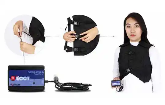
Principally, the ECCT consists of two parts: the apparel as a support of the capacitive electrodes and oscillator to generate electric wave with certain intensity, waveform and frequency.
The ECCT specification for treating cancer is determined by the coverage of the apparel, the frequency, the intensity and waveform of the oscillator, and the time of usage of the equipment that correlate to time exposure of the cancer to electrostatic wave.
For complete removal of the cancer, the apparel design is essential in the treatment method, and must be customized according to the tumor position and its staging. In principle, the coverage of the apparel is divided into two types: global coverage for metastasize prevention and local customized coverage for total destruction of the primary tumor.
The frequency, intensity and waveform of the oscillator, and the time of usage are determined based on the grade of malignancy of the cancer, the pathology anatomy and the electric properties of the cancer cells. In general, the higher the level of the staging and the higher the degree of the malignancy the more responsive of the cancer to the electric wave and thus the less time needed for exposure as the body has limited capacity to absorb and dissolve the resulted dead cells.
Process and Progress of Cancer Treatment with ECCT
Figure 2 shows the macroscopic process of “melting” cancer cells and regenerating normal cells during the treatment of already open wounded breast cancer. The figure shows that the destruction of cancer cells occur in very short time of within a month, while the regeneration of the normal cells depends on the total removal or absorption of the dead cells and could take years if conducted naturally.
A. Initial treatment
B. 2 months of treatment
C. 3 months of treatment
D. 4 months of treatment
E. 5 months of treatment
F. 2 years of treatment
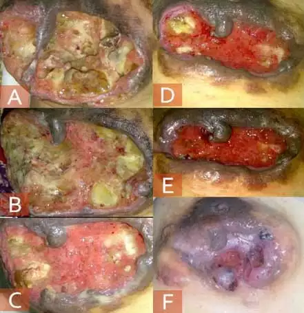
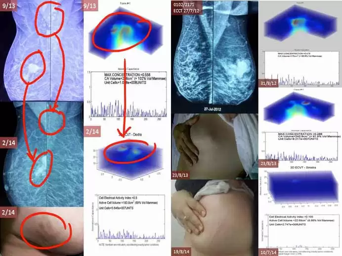
Figure 3 shows two cases of images of the breasts taken by mammography and electrical property
imaging using ECVT (Electrical Capacitance Volume Tomography, Taruno et al., 2014) during ECCT treatment. The mammograms on the left shows disappearing small axillary nodules after 6 months of ECCT treatment and dissolving relatively big size (>2 cm in diameter) primary nodule on the breast, while the photograph (left) shows darkened skin as a result of the discharged dead cells. The right photograph shows that the relatively big lump eventually disappear after 2 years of treatment with complete discharge of the dead cells. The ECVT images shows gradual decreases in the electrical properties of the tumors in both cases with the treatment.
Why are healthy cells unaffected by ECCT?
The electric properties in cancer cells are different from the healthy ones. Cancer cells have relatively higher electric properties (conductivity and permittivity) as compared to normal cells. Consequently, cancer cells are relatively more responsive to external electric fields than normal cells.
The response of cancer cells to external electric field is more salient and destructive during the process of cell division due to the high electric tension generated by micro-tubule activity during the mitosis process. The ECCT is set low enough in the intensity and the frequency to relatively only affect the cancer cells during mitosis.
How to remove dead cancer cells?
A proper ECCT exposure to the designated location of tumors can destroy and break down the cancer cells in relatively short times in the range of few days to few weeks. If designed and used properly, ECCT can exterminate a cancer lump of few centimeter in size within weeks. However some issues may arise to the dead cancer cells if accumulated beyond the capability of the body to absorb and excrete to outside the body in the form of body excretions.
The dead cancer cells contain 70% water, 20% protein, and the rest of gas. If the metabolism of the patient is normal, the dead cells can usually be easily absorbed by the blood and discharged through urine, feces, sweat, or phlegm that sometimes comes out with extremely bad odor as a result from decomposed proteins.
The effectiveness of the treatment to completely kill the cancer cells will depend on the ability of the body to absorb and resolve all the dead cells as the dead cells, if accumulated, could cause depolarization of the electric field and prevent further process of treatment. Surgery is, therefore, mostly recommended to remove completely and safely the dead cells. For the case of metastasized cancers, the effectiveness of the treatment depends on the sizes of the metastasized lumps, but not the degree of the spread. Small lumps or nodules regardless of the extension of the distribution in the body is relatively easy to be destroyed by the technique, and the resulted dead cells is easily absorbed and disposed by the body.
Therefore the technique is most effective to be used in conjunction with surgery, to clean and prevent metastasize before and after surgery.
Procedure of ECCT Treatment
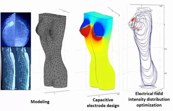
The procedure of the ECCT treatment comprises three steps:
- Localizing the tumor based on the MRI or CT scan images
- Designing capacitive electrodes and computing of the electric field distribution inside the treating domain of the tumor, and
- Optimizing the electrode design based on the electric field intensity distribution to attain enough intensity required in the tumor site by considering the possible discharge channel to dispose the dead cells through connected veins linked with the tumor location.
Figure 4 Three steps of ECCT treatment stepwise: (1) Finite element method modeling based on the location of the tumor in MRI or CT scan images, (2) Electrode design and electric field computation, and (3) Optimization of required electric field intensity distribution.
Hence, Understanding the suitability of the device for your specific condition is paramount, we emphasize a structured process. Only through a proper study of your medical condition can we recommend the most suitable ECCT devices for you.
The initial step involves gathering comprehensive information about your medical condition. We kindly request the following medical reports to facilitate the preparation of a personalized use case report:
- Recent Scans (MRI, CT, etc.): Please provide any recent imaging scans that can offer insights into the current state of your condition.
- Doctor’s Treatment History: We also require a detailed treatment history from your healthcare provider, including past and current treatments, medications, and any relevant observations.
Once we receive these documents, our team of experts will conduct a thorough analysis to generate a use case report called ECCT Assessment Report Book yours today. This report will outline the recommended ECCT devices suitable for you based on your specific condition.
The Differences Between ECCT and Tumor Treating Fields (TTF)
Electro-Capacitive Cancer Therapy (ECCT) and Tumor Treating Fields (TTFields) are devices that used to treat cancer based on alternating current (AC) electric fields.
The ECCT itself comprises of two parts, the oscillator and the apparel. ECCT oscillator is a box-shaped component used for generating electric fields when it is connected to ECCT apparel. It has some voltage and frequency types. Its complete specifications are written down in Table 1. ECCT apparel is a device in the form of clothes worn by cancer patients. Its complete specifications are written down in Table 2.
- Charging Adapter
- Charging Input
- Battery Type
- Charging Time (internal)*
- Charging Time (external)*
- Dimension
- Weight
- Connector Type (Charging)
- DC 5V, max. 500mA
- AC100-240 50/60Hz
- AAx2 Ni-MH>2000mAh
- 3 – 7 Hours (optional)*
- 2 – 3.5 Hours
- 80mm x 50mm x 30mm
- Approx 90g
- USB Mini type B
Table 1. ECCT oscillator specifications
- Outer fabric
- Dacron
- Nonwoven
- 70% Polyester 10% Polyamide Interlining
- Dacron
- Nonwoven Kufner
Table 2. ECCT apparel specifications
For treating cancer, ECCT uses low-intensity electrostatic wave source (18-30 Vpp) and intermediate frequencies (100-200 KHz) that produces electric polarization on the near field area which is bordered by several capacitive electrodes installed in the apparel. ECCT generates electric fields from the non-contact capacitive electrodes which are placed around the tumor, with the exact frequency and intensity to disturb the microtubule polymerization process that can inhibit cell division, so that the cancer cells eventually die. With the intermediate frequency and low intensity, ECCT is basically safe, relatively has no side effect and it does not disturb the normal cells.
There are 3 main differences between ECCT and TTFields
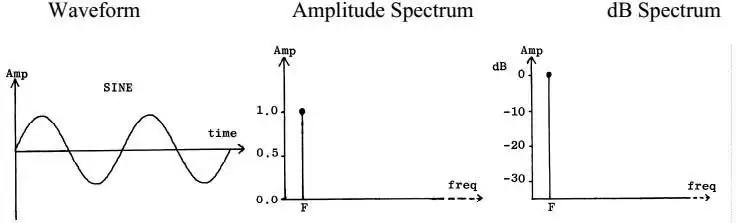
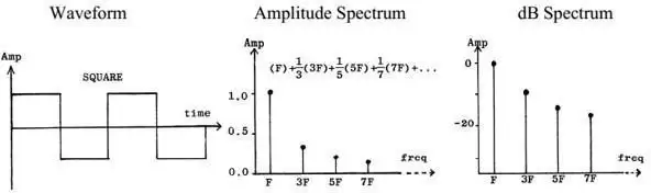
The waveform of electric fields
TTFields exerts sinusoidal electric waveform or better known as Sine wave (Fig. 1). While, ECCT exerts square electric waveform from an oscillator (Fig. 2).
The contact between electrodes of the device into the surface of the body
TTFields uses electrodes that directly contact to patient’s body (Fig. 3). This direct contact may cause dermatitis beneath the electrode gel as reported by Kirson et al. (2007). While, ECCT uses non-contact electrodes that do not make a direct contact to patient’s body (Fig. 4).
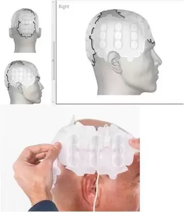

Figure 3. Contact electrodes of TTF on a head and body of a patient.


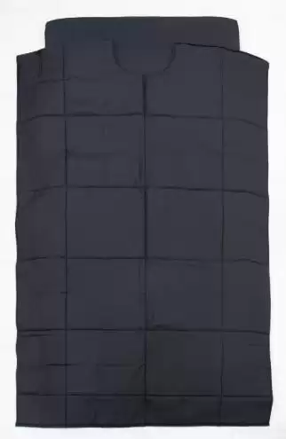
Figure 4. Non-contact electrodes inside the apparels of ECCT
The source of electricity
ECCT uses small battery inside the oscillator (Fig. 4); while TTF uses big battery as a TTF generator (Fig. 5)
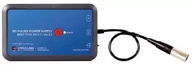

Figure 4. A battery as an electric source for TTFileds.
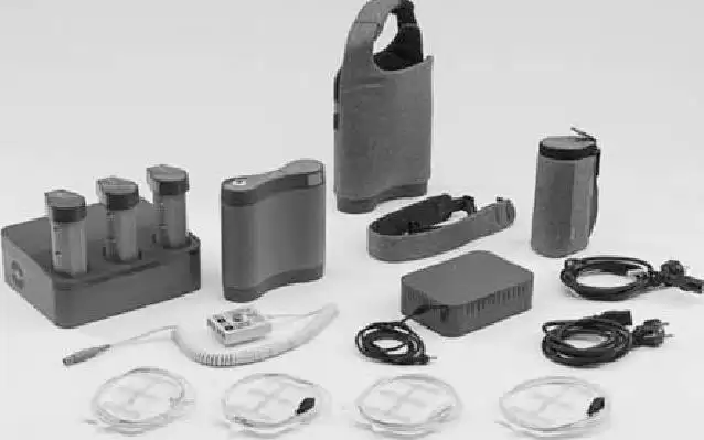

Figure 5. A battery as an electric source for TTFileds
How ECCT differs from TTF (Optune Co.) Method?
ECCT (Electro-Capacitive Cancer Therapy)
 Capacitive (pure electric field, no current);Completely Non-contact;Penetrating through air layers;Relatively low voltages (<+-30Vpp)Effective to air-contact or interfacial tumors (not so good to bulk solid tumor)
Tumor Treating Fields (TTF)
Quasi -Electrostatic (quasi-conductive, electric field containing current)Using high-conductive ceramic electrodes with direct contact to skinNo -air layer permittedRelatively higher voltages (>50V)Work only to solid tumor (Might not possible for air-contact tumor)
Routine Monitoring and Consultations
ECCT treatment is highly customized according to the patient’s conditions, the cancer staging, the pathology anatomy and its response to electric field. All treatment is done daily at home while conducting daily activities. The present of doctors or medical physic therapist for routine consultation during the process of treatment is essential for effective customized and safe treatment to achieve complete cure, which is possible with the ECCT even in the late state of cancers.
Routine Checking of Devices
ECCT is used every day and for a long period of time. Thus, it is necessary to keep up a routine checking (once a month) of ECCT equpments to ensure that all the devices work properly.
- Mursilatun, Electric field effect on growth of cancer cells. Bachelor of Thesis, 2010, Dept. of Medical Physics, University of Indonesia.
- Sabrina, Qolby, Effect of electric field treatment variation against Lethal level cell line MCF-7 (human breast cancer) in vitro and measurement of cell capacitance value. Master Thesis, 2014, Dept. of Medical Physics, University of Indonesia.
- Ajrina, Izzatun, In vitro trial on antiproliferative and cytotoxic effect of 100 KHz electric field towards MCF-7 breast cancer cells. Bachelor Thesis, 2013, Dept. of Biology, University of Airlangga.
- Salim, Sahudi, The electro-static field effect on growth of Hella cell (human epithelial carcinoma cell line), an in vitro study. Dissertation, 2015, School of Medicines, University of Airlangga.
- Handayani, Yunita Kusuma, The effectiveness of Electro-Capacitive Cancer Therapy (ECCT) in breast cancer treatment. Bachelor Thesis, 2012, Dept. of Medical Physics, University of Indonesia.
- Amdanita, Putri, Evaluation of capacitive electrode design to improve effectiveness of Electro-Capacitive Cancer Therapy (ECCT) for breast cancer treatment. Bachelor Thesis, 2013, Dept. of Medical Physics, University of Indonesia.
- Nurzannah, Analysis of Electro Capacitive Cancer Treatment (ECCT) Efficiency in Breast Cancer Treatment Based on breast Anatomy. Bachelor Thesis, 2013, Dept. of Medical Physics, University of Indonesia.
- Nurhasanah, Siti, Electrode design optimization for Electro-Capacitive Cancer Therapy (ECCT) in treatment of breast cancer. Bachelor Thesis, 2013, Dept. of Biomedical Physics, Bandung Institute of Technology.
- Hendriyanto, Markus, Effectiveness of Electro-Capacitive Cancer Therapy (ECCT) for brain cancer treatment. Bachelor Thesis, 2013, Dept. of Medical Physics, University of Indonesia.
- Yulianto, Ahmad, Electro-Capacitive Cancer Therapy (ECCT) design optimization for treatment of carcinoma Nasopharing. Bachelor Thesis, 2012, Dept. of Medical Physics, University of Indonesia.
- Dimyati, Hamid and Sri Haryatmi: “Weibull Regression model for testing factors affecting survival of cancer therapy (ECCT)”, Proc. of ICAS 2014, Khun Kuen, Thailand.
- Taruno, Warsito P., Marlin R. Baidillah, Rommy I. Sulaiman, Ifnia Widora, Arbai Yusuf, Wahyu Widada, Muhammad S. Aljohani, Frans X. Suharyanto: “Comparission of Electrical Capacitive Volume Tomography and ultrasonography for breast cancer detection”, Advance Science, Engineering and Medicine, Volume 6, 2014, pp.845-848 (4).
Copyright by CTech Labs Edwar Technology, 2015
- https://pubmed.ncbi.nlm.nih.gov/32695310/
- https://pubmed.ncbi.nlm.nih.gov/34164110/
- https://iopscience.iop.org/article/10.1088/1742-6596/1863/1/012036/meta
- https://karger.com/imi/article/4/3-4/161/176180/Cell-Death-and-Induced-p53-Expression-in-Oral
- http://www.embj.org/embj/eletric-field-distribution-cancer-treatment/